en | es | val
en · es · val

Breast cancer

diagnosis · treatment · research

What is breast cancer

Breast cancer is a disease in which breast cells multiply uncontrollably, becoming cancerous and resulting in the appearance of a mass of transformed cells (tumour), with abnormal growth and multiplication.

However, in order to understand what breast cancer is, it is useful to delve a little deeper into the anatomy of the female breast. The breast is made up of lobes, which in turn are divided into smaller structures called lobules. The lobules are responsible for producing milk when a woman is breastfeeding. This milk travels from the lobule to the nipple through tubes called ducts.

Additionally, as with all parts of the body, the breasts have lymphatic vessels that connect to small, round organs called lymph nodes. The lymph nodes are responsible for protecting our body from anything they consider foreign: they trap tumour cells, bacteria and other harmful substances.

Breast cancer, as we mostly understand it, is a disease that originates in the mammary gland, specifically in the ducts or tubes that carry milk to the nipple, or in the lobules, which are the glands that produce milk. It is a disease that mainly affects women, although it can also occur in men.

The type of breast cancer will depend on which cells in the breast become cancerous. But broadly speaking, there are two types of breast cancer:

In situ or non-invasive breast cancer. In this type of cancer, the tumour cells are in the ducts or lobules of the breast, but have not spread to the rest of the healthy breast tissue. It is, therefore, a tumour with a better prognosis than invasive tumours.

Invasive breast cancer. In its evolution, breast cancer can grow locally, invading other parts of the breast or neighbouring structures such as, for example, the chest wall and the skin. On the other hand, when there is lymphatic spread, the cancer spreads through the lymphatic vessels to the armpit, or through the blood vessels in the breast, reaching other organs such as the bones, lungs or liver. When this occurs, it is called metastasis.

Depending on the progression and severity of the disease, breast cancer is also classified into:

  • Early-stage breast cancer. This is diagnosed when the tumour is confined to the breast or axillary lymph nodes.
  • Locally advanced breast cancer. It is diagnosed when the cancer invades tissues close to the breast or lymph nodes.
  • Metastatic breast cancer. When breast cancer spreads to other parts of the body such as the bones, lungs or liver it is called metastatic, which is the breast cancer with the most serious prognosis.

Breast cancer staging is used to describe its size, extent, and whether it has spread from the organ where it originated. There are five stages of breast cancer (0 to IV), and the higher the stage, the worse the prognosis.

Breast cancer staging is used to describe the size of the cancer, its extent, and whether it has spread from the organ where it originated. For breast cancer, there are five stages (0 to IV) and prognosis worsens the higher the stage.

  • Stage 0. Non-invasive or in situ tumour, located in the breast. The tumour is small and its location is limited to the breast tissue. This stage is further subdivided into two:
  • Stage II. The tumour may be in the breast and/or lymph nodes.
  • Stage III. The tumour has spread from the breast to lymph nodes near the breast, to the skin of the breast, or to the chest wall.
  • Stage IV. This is the worst prognostic stage and is diagnosed when the tumour has spread distantly to other parts or organs of the human body.

Epidemiology of breast cancer

33.375

33,375 new cases of breast cancer were diagnosed in Spain in 2021

86%

The survival rate of women with breast cancer after 5 years is 86%

1 out of every 8

The estimated probability of developing breast cancer is 1 in 8

Causes and symptoms

The causes of breast cancer and why it arises are currently unknown, but we do know about the so-called risk factors, i.e. situations that can increase the likelihood of developing breast cancer.

It is worth remembering that having one or more risk factors does not mean that you will get cancer in the future. Similarly, not having a risk factor does not mean that you can never get breast cancer.

  • Being a woman over 50
  • Genetic predisposition with a family history (genes involved in DNA repair mechanisms such as BRCA1 and BRCA2)
  • Previous exposure to ionising radiation
  • Reproductive history
  • Breast density
  • Alcohol consumption
  • Obesity

The most common symptoms are:

  • the appearance of a lump in the breast or armpit
  • changes in the skin of the breast
  • nipple retraction

If any of these symptoms appear, it is a priority to see a doctor or specialist. Breast cancer is a tumour that can be diagnosed early, even before the patient detects any symptoms, and this is possible thanks to early diagnosis by mammography. This mammography is recommended for women between the ages of 45-50 years (in the Valencian Community, at 45 years of age). The Instituto Valenciano de Oncología Foundation has had an Early Diagnosis Unit since 1982, where mammography screening is carried out to detect breast cancer.

The Importance of Early Diagnosis

Breast cancer is a tumour that can be diagnosed early, even before the patient detects any symptoms, and this is possible thanks to early diagnosis by mammography.

The IVO has had an Early Diagnosis Unit since 1982, where mammography screening is carried out to detect breast cancer. You can contact the unit directly to make an appointment. 

How it is diagnosed

Breast cancer is one of the few cancers that can be detected early by minimally invasive tests. The well-known mammography has been credited as the best and most effective screening test for breast cancer. However, in the detection and diagnosis of breast cancer, the process is as follows:

The first important test for diagnosis is regular physical examination, both by the patient and by a physician.

If there is a suspicion of a lump or other symptoms associated with breast cancer, such as changes in the skin of the breast or nipple retraction, the radiological diagnostic process would be initiated in order to perform a breast imaging examination.

If there is a suspicion of neoplasia or cancer after the physical examination, it will be necessary to carry out a mammography, which we could say is an X-ray of the breast.

The IVO has a breast cancer early diagnosis unit with more than 40 years of experience in diagnosis.

To find out more precisely whether the lump or tumour detected is solid or a fluid-filled cyst, an ultrasound (a technology that uses high-frequency sound waves to create an image of the interior) will be performed. Finally, if the above tests are inconclusive, an MRI scan, which uses magnetic fields and radio waves to produce detailed images, would be performed. An MRI can detect the presence of multiple tumours.

The next step is to take a sample from the lesion. In most cases, a core needle biopsy (CNB) will be performed, as it allows a larger amount of tissue to be obtained than a fine needle aspiration (FNA) and therefore provides a more accurate diagnosis. In both cases, a sample of cells or breast tissue is obtained to be studied under a microscope by a specialist in anatomical pathology.

A biopsy involves the collection of tissue located anywhere in the body for subsequent examination under a microscope to determine the presence of a disease. Sometimes, depending on the extent and suspected diagnosis, a biopsy may be performed to remove a small sample of tissue with a needle, while in other situations a biopsy may involve the complete resection of a suspicious lump or nodule. In each case, the medical professional performing the biopsy will give instructions to the patient according to the type of biopsy to be performed.

In cases of breast cancer, a lymph node biopsy is a very important aspect, as it allows for the staging of breast cancer and is performed to confirm or exclude tumour involvement/extension to the axillary lymph nodes.

The definitive diagnosis of breast cancer is made after observing the malignant cells under the microscope, and is established by the specialist in anatomical pathology. As previously mentioned, the pathologist identifies the origin of the tumour cells: ductal, lobular or other; their invasive capacity: infiltrating or non-infiltrating tumour; the histological grade; the expression or not of hormone receptors, oestrogen and progesterone; and the expression or not of the HER2 protein.

The pathologist will issue a report explaining all these aspects, which will be crucial in deciding on the best therapeutic option for the patient.
Most diagnostic techniques are currently carried out by the pathologist. On special occasions, other techniques for analysing mutations in certain genes are performed by the Molecular Biology service. Such techniques are mainly performed in metastatic breast cancers, and the identification of mutations in specific genes will allow us to offer targeted treatments against the detected mutation, which is what we call personalised treatment.

With all the information obtained—from the interview with the patient to learn about their medical history and symptoms, from the physical examination, from the results of the imaging techniques, and from the pathologist’s report—the IVO breast cancer medical staff and members of the Breast Cancer Tumour Committee, both together and after evaluating each case individually, will offer the patient the best therapeutic option as well as possible alternatives.

Hereditary breast cancer

It is estimated that only between 5 % and 10 % of all cancers are hereditary

Breast cancer, ovarian cancer and colorectal cancer are the tumours most frequently associated with hereditary syndromes.

The aim of the Genetic Counselling Unit is to identify people who carry a pathogenic mutation in genes that are currently known to be associated with an increased risk of developing one or more types of neoplasms. In many cases, these people have a family history suggestive of hereditary transmission. Although they have no clinical signs of suffering from cancer, they present a high probability of developing the disease some time in their lives. Generally, the specialist doctor will refer the patient or family member to the Cancer Genetic Counselling Unit (UCGV, for the Spanish acronym) to complete the study.

According to international clinical practice guidelines, it is recommended that genetic testing be offered when:

  • The individual has a personal or family history suggestive of an inherited cancer predisposition syndrome
  • The results of the genetic study can be interpreted
  • The results of the genetic study will aid in diagnosing or influencing the medical or surgical management of the individual or their at-risk relatives.

Treatment of breast cancer

The treatment of patients with breast cancer depends on many factors, but it is mainly based on the extent of the disease (known as the “stage”), the type of breast cancer according to the anatomical pathology report, and the patient’s risk assessment.

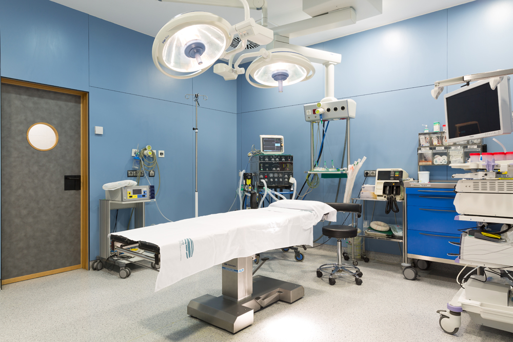
In the case of breast cancer, the aim of surgery is to remove the tumour and analyse the lymph nodes in the armpit (lymphadenectomy), always with the premise of opting for the least aggressive surgical technique adapted to the individual. Basically, there are two main types of surgery for women with breast cancer:

  • Breast-conserving surgery and mastectomy. Breast-conserving surgery is the least aggressive technique, as a partial removal of the breast is performed and the breast is not completely resected, unlike a mastectomy. The tumour is removed, with safe surgical margins and then local or loco-regional radiotherapy is administered to eliminate any tumour cells that may remain after the surgery.
  • Mastectomy. In this type of surgery, the entire breast is removed, which may or may not include the nipple-areola complex. During the same surgery, it is possible to reconstruct the breast (immediate reconstruction), or it can be done once the complementary treatments have been completed (deferred reconstruction).

One of the first sites where breast cancer spreads is to the lymph nodes in the armpit. The extent to which this occurs will determine the prognosis of the breast cancer. The only way to know if breast cancer has spread to these lymph nodes in the armpit is to examine them under the microscope. To do this, some nodes have to be removed and then analysed. This procedure has side effects that can be very uncomfortable, such as temporary limitation of arm movement and numbness or swelling of the arm (also known as lymphoedema).

In a specific group of patients, especially those in whom there is clinically no tumour involvement of the sentinel nodes, an alternative to lymphadenectomy may be sentinel lymph node biopsy. The sentinel node is the first node where the tumour is likely to spread. The procedure involves injecting a radioactive substance near the tumour that will flow through the lymphatic ducts to the lymph nodes, detecting which lymph node is the first to receive the substance and testing it after removal. If the result is negative, the lymphadenectomy is no longer necessary and the patient can avoid the side effects described above.

Radiation therapy is a type of treatment that uses ionising radiation to destroy tumour cells and prevent their growth. It damages the DNA of these cells, causing them to die. It is used as a complementary treatment to surgery. It is usually performed after conservative surgery or, in some cases, after a mastectomy.

Radiation therapy can be performed on the breast, chest wall and/or loco-regional lymph node areas such as the axilla or supraclavicular level. The usual form of administration is external radiation therapy, administered by means of a machine (linear accelerator). The duration of treatment can vary from days to weeks, depending on different factors, and just like with surgery, effective strategies with the shortest possible duration and with the least possible associated side effects will always be sought out.

As a complementary treatment to external radiation therapy or as an alternative in some cases, internal radiotherapy or brachytherapy is also used, which consists of injecting radioactive material into the area of the tumour.

Chemotherapy is the administration of drugs, usually intravenously, with the aim of destroying tumour cells scattered throughout the body that surgery and radiation therapy cannot attack. It is described as an adjuvant treatment when administered after surgery, whereas it is called neoadjuvant chemotherapy if chemotherapy is used to shrink the tumour before surgery. The decision to administer the treatment before or after surgery will be made by the multidisciplinary committee and then agreed upon with the patient’s consent.

Chemotherapy treatment is administered in cycles, and the duration and number of cycles will depend on each case. Each cycle lasts on average between once a week or every 2-4 weeks, and the reason why a chemotherapy treatment is administered in cycles is due to the effectiveness of the treatment itself: the treatment is administered in cycles in order to attack the cancer cells when they are most vulnerable and to give the body’s healthy, normal cells time to recover from the damage suffered by the treatment.

Chemotherapy usually causes a series of side effects, including fatigue, nausea and vomiting and temporary hair loss, among others. As with local therapies (surgery and radiotherapy), chemotherapy is intended to be administered only to those patients where the potential benefits, in terms of reducing the risk of recurrence, outweigh the side effects. The current use of genomic platforms can reduce the administration of chemotherapy in up to 40% of cases. In our centre, it is possible to request this determination in those cases in which it is indicated.

Oestrogen and progesterone are sex hormones that are naturally present in women. As previously discussed, some breast tumours depend on the supply of these two substances to grow, and as many as 70% of breast cancers do. When tumour cells retain these hormone receptors, it is possible to administer a drug or hormone treatment that has been shown to reduce the risk of tumour relapse and improve survival rates. Hormone therapy can be administered orally and has fewer side effects than other treatments, such as chemotherapy. The duration of this treatment may range from 5 to 10 years. Monitoring of possible side effects is carried out by the medical oncologist, so the patient will attend quarterly or semi-annual check-ups to find out about possible side effects, as well as any symptoms related to other treatments or those that could lead to the suspicion of a possible tumour recurrence.

Between 15-20% of breast cancers present with an overexpression of the HER2 protein, and these are known as HER2-positive tumours. HER2 proteins are found on the surface of breast cells. HER2 is a transmembrane protein, which spans the entirety of the cell membrane and is involved in normal cell growth. When the tumour has a high amount of this protein, the cancer tends to spread faster than other types of breast cancer. There are now drugs known as monoclonal antibodies (trastuzumab and pertuzumab), which act on tumour cells that can overproduce the HER2 protein, stopping or reducing their growth. In other words, these drugs have the potential to change the nature of this subtype of breast cancer and turn a poor prognosis to a good prognosis due to their high efficacy. These agents are usually administered in combination with chemotherapy for twelve months.

Treatment of breast cancer at the IVO

Breast cancer is one of the few cancers that can be detected early by minimally invasive tests. The well-known mammography has been credited as the best and most effective screening test for breast cancer. However, in the detection and diagnosis of breast cancer, the process is as follows:

IVO Breast Tumour Committee

The Breast Tumour Committee is made up of a multidisciplinary team of expert professionals.

Medical oncology service

The Service's team of professionals accompanies the oncology patient throughout the disease process.

General surgery service

The breast section of the surgery service is responsible for surgery in patients with breast cancer.

Clinical trials

The only way we currently have to advance and improve cancer treatment is through so-called clinical trials.

A clinical trial is a research study carried out on people with the aim of learning more about how the body reacts to certain treatments. These trials generally seek to find drugs that are more effective than the current best therapeutic option for patients, or that have similar efficacy but a better toxicity profile.

Bearing in mind that almost all currently available treatments are the result of clinical research, the importance of clinical trials is obvious.

The IVO has a clinical trials unit for all types of tumours and participates in phase 1-3 studies as well as other types of studies. To date, we have participated in more than 100 studies for breast cancer, some of which were designed by our own researchers and have become the new therapeutic option in different types of breast cancer such as luminal, triple negative or HER2 positive.

Adverse effects

Each type of cancer will require a specific treatment, and the adverse effects will largely depend on the treatment applied to each patient. These will be monitored and managed by the relevant specialist. However, there are common adverse affects for each type of treatment.

The adverse effects of any anticancer therapy are graded from 0 to 5, with 0 meaning no adverse effects, 1 being the mildest , 2 being moderate, 3 being severe, 4 being life-threatening, and 5 being death due to the treatment.

The goal is to identify the grade of each adverse effect that the patient may be experiencing in order to minimise them. To do so, some decisions may be made, such as maintaining treatment at the same doses, stopping the treatment temporarily, reducing the dose, or permanently discontinuing the treatment.


After breast cancer surgery, a high percentage of patients suffer from what is known as lymphoedema. Lymphoedema or lymphatic oedema is an after-effect of breast cancer treatment. It occurs when the arm by the affected breast swells due to fluid accumulation. This is because breast cancer surgery often involves the removal of lymph nodes in the armpit, making it difficult for lymph to be eliminated, causing this fluid to accumulate in the arm and producing oedema.

If lymphedema occurs, it is advisable to be extremely hygienic with the skin of the affected arm, avoid tight-fitting clothes on the shoulder and arm, avoid taking blood samples and blood pressure tests in that arm, not apply excessive heat to the area, not carry weight on that arm, and not wear watches or bracelets. In addition, it is recommended to do some exercises to help maintain the arm’s mobility. The specialised physiotherapist will help and guide the patient in cases of lymphoedema, for which a drainage treatment or lymphatic massage and compression bandage can be applied, always under their supervision.

There are also some common adverse effects of radiation therapy, such as pain and swelling in the breast where the radiation is administered, fatigue and skin irritation. Your doctor will advise you on how to manage these side effects, but using creams can help with irritation, as can avoiding exposure to sunlight.
The adverse effects of chemotherapy for breast cancer vary depending on the amount, type and dose of the drugs used. Each patient should discuss with their doctor what these will be in each case.

In general, tiredness or fatigue has been described as one of the most common adverse effects. This can be caused by the cancer itself or by the treatment. The recommendation will always depend on the doctor or specialist treating you. It is always advisable to consult with the medical staff, who will help you to better cope with this tiredness. Also, nausea and vomiting are common in patients receiving chemotherapy, and the development of new drugs makes it possible to control symptoms in more than 80% of cases. Finally, it should be noted that most of the adverse effects of chemotherapy for breast cancer are mostly temporary and can be controlled with medication, and specialised medical staff will help you to deal with them.

Living with breast cancer

coping with the diagnosis, receiving treatment, breast reconstruction, psychological support and regular check-ups

Whether you receive the news of an initial diagnosis of breast cancer or a relapse, coping with breast cancer can be emotionally overwhelming. Each person has their own way of coping with a breast cancer diagnosis, but there are some recommendations that can help you through this process:

  • Maintain communication and the company of family and friends, the people closest to the patient, who can provide a support network throughout the process
  • Talk to other people who have survived breast cancer or who are in the same situation. There are many local and national associations and support groups. The Spanish Association Against Cancer (Asociación Española Contra el Cáncer) is perhaps the best-known one.
  • Inform yourself in order to make the best decisions about treatment and medical professionals.

Guide for breast cancer patients

To provide further information, a multidisciplinary team of professionals from the IVO Foundation has produced a complete guide to breast cancer, which can be downloaded below in PDF format. 

diagnóstico precoz

Medicina nuclear

CIRUGÍA ROBÓTICA

radioterapia