


The Pneumology Unit deals with the study, diagnosis and treatment of respiratory pathologies that oncology patients treated at the center may present and aggravate with their tumor process, as well as pulmonary involvement secondary to the neoplastic disease and the different treatments (surgery, radiotherapy, chemotherapy, immunotherapy and other targeted therapies). All of this in close collaboration with other services such as Internal Medicine, Thoracic Surgery, Oncology, etc.
Special dedication to the early diagnosis of lung cancer in people at high risk of developing it. Since 2008 we have been developing a screening program with low-dose radiation CT; integrated by a multidisciplinary team of radiologists, interventional radiologists, pulmonologist, thoracic surgeons, pathologists and oncologists.
Activity of the unit:
– Respiratory functional assessment of the oncologic patient, necessary prior to lung resection surgeries, radiotherapeutic treatments and others.
– Diagnostic techniques such as bronchoscopy for histological or microbiological sampling, both in the diagnosis of lung cancer and other concurrent processes.
– Study of solitary pulmonary nodule.
– Lung screening focused on the early diagnosis of lung cancer. It is a follow-up program with low-dose radiation CT aimed at patients at high risk of developing it.
– Smoking cessation, the main agent causing respiratory pathology.
– Support in the diagnosis – treatment and management of pulmonary pathology, to other hospital areas such as Emergency, Hospitalization and home hospitalization.
– Management of infections, pulmonary toxicities or venous thromboembolic disease, pathologies especially prevalent in oncology patients.
The Pneumology Unit collaborates with the I-ELCAP Early Detection Unit, where they perform the early diagnosis of asymptomatic lung cancer
Early Diagnosis of Asymptomatic Lung Cancer
Lung cancer is one of the most common cancers and the one that causes the highest mortality (higher than breast, cervical, colon and prostate cancers combined). Its prognosis is related to the stage of the disease at the time of diagnosis.
Tobacco is the main cause of lung cancer. It also causes other pulmonary pathologies such as emphysema, which independently increases the risk. Smoking cessation is the most effective measure to reduce it.
Screening makes it possible to detect most types of lung cancer in early stages, before the patient presents symptoms, when it is possible to apply a curative treatment, thus reducing mortality.
It includes people between 55 and 70 years of age, without alarm symptoms but at risk of developing lung cancer because they are or have been smokers. Other risk factors such as emphysema, EPOC, contact with asbestos, first-degree family history of lung cancer, etc. are considered.
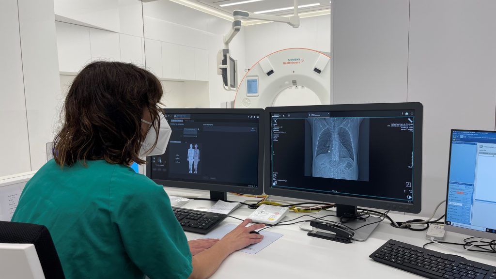
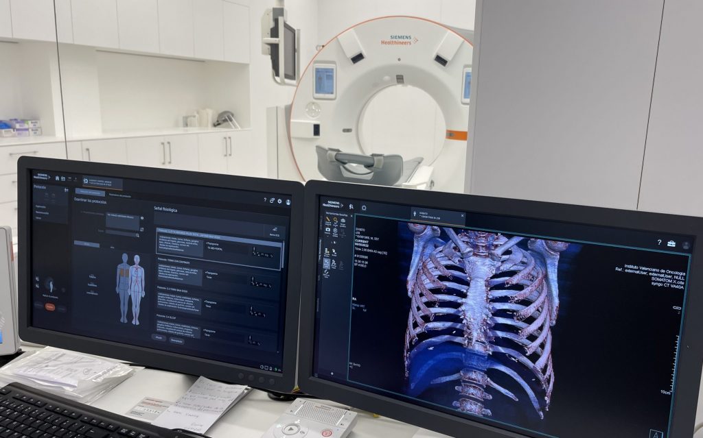
We use low-dose radiation computed tomography (TCBD) to detect suspicious lesions; depending on their characteristics, size, etc., follow-up is decided. The radiation dose is about 0.8 mSV, similar to that of a mammogram and about seven to ten times lower than that of a standard CT scan.
The IVO offers you a screening with a time-limited follow-up, during which we will inform you of any findings and, if necessary, perform the diagnostic study.
Diagnostic Techniques
Respiratory Function Tests
A technique that allows, through simple respiratory manoeuvres, for the measurement of pulmonary functional capacity and the detection of alterations that are secondary to various diseases (asthma, bronchitis, emphysema, fibrosis, tumours, etc.).
Fibrobronchoscope
It uses an instrument called a fibrobronchoscope (a long, narrow, flexible tube that uses fibre-optics). This device allows for the direct exploration of the anatomy of the larynx and the tracheobronchial tree, as well as taking biopsies from possible lesions within the lungs. The procedure is usually performed under a local anaesthetic and sedation or, at times, under general anaesthesia.
EBUS/EUS (Endobronchial Ultrasound/Oesophageal Endoscopic Ultrasound)
Similar to the technique above in which the fibrobronchoscope carries a small ultrasound scanner in its tip that allows the user to view and puncture structures that externally surround the tracheobronchial tree and oesophagus.
Pneumology Service Medical Team
Associated Doctor
Dr. Encarna Martínez Pérez