

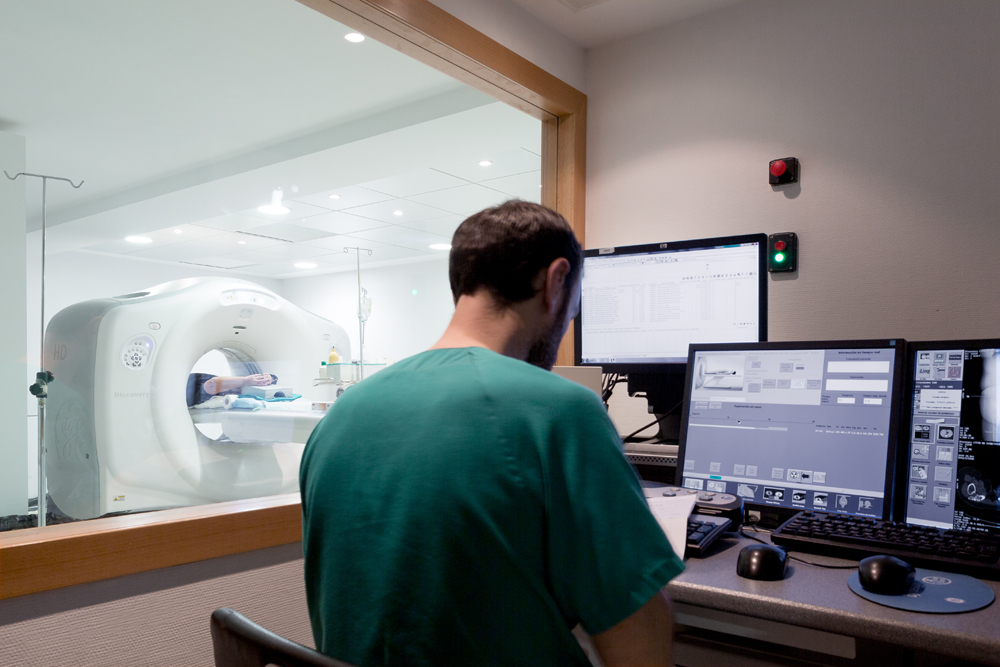
To correctly treat an oncological pathology, it must first be properly diagnosed and its staging must be precisely defined. The radiological studies that are performed in the Radiology Service allow for the diagnosis of tumour pathology as well as monitoring the disease and its response to the treatments applied, making this department essential in the oncological process.
The Radiology Service at the IVO is staffed by a team of specialists with extensive and proven experience in oncological pathology, distributed by care areas in relation to the type of tumour.

The IVO’s Radiology Service participates in the corresponding hospital’s Tumour Committees
Areas and activity of the service
- Vascular Radiology
- Breast Pathology
- Technological Innovation
- Collaboration
- Continual Professional Development
- Research
- File and Management System
- Early Cancer Diagnosis
The service contains the Vascular and Interventional Radiology Area, which carries out any type of image-guided diagnostic or therapeutic procedure, from minimally invasive treatments to advanced tumour chemoembolization and tumour ablation by inserting selective devices.
Diagnostic-therapeutic management of breast pathologies requires a radiology area with experts in interpreting these images and the types of tumours. At the IVO, they have all the necessary technology for attending to this pathology: 3D digital mammography with tomosynthesis, magnetic resonance, ultrasound, prone table and biopsy techniques.
Technological innovation in the field of radiology facilitates and enhances the visualisation of lesions, offers a higher-quality image, allows for earlier detection of the disease, lowers the dosage of radiation to be used in treatment, increases the speed and, therefore, provides greater diagnostic reliability.
This Service works closely with the Radiophysics Service to ensure good image quality and the safety and welfare of the patient by controlling radiation during the examination, both in the diagnostic tests and while monitoring the disease.
Participation in the hospital’s Tumour Committees, attendance in scientific meetings, specialisation in new diagnostic techniques, sharing information with other cancer specialists, and continual professional development allow the practitioners that make up the Radiology Service to have a broad, current and dynamic knowledge of the advancements in oncology.
In terms of the research activity, the service takes part in the various research studies and clinical trials for therapeutic evaluation aimed at cancer treatment.
The IVO has a powerful file, management, and communication system for radiological images and their corresponding reports called PACS, where all patient images are integrated. These images and diagnostic tests are digitised and can be accessed by the hospital’s physicians, which keeps all the information centralised in one place, allowing for the precise monitoring of a disease’s evolution and its response to treatment. For patient monitoring, the application of functional imaging techniques using computer programs helps to assess the response to the therapy used.
Early diagnosis to detect lung cancer in its early stages is very important, since it allows for either the possibility of surgical treatment or a combination of therapies that are less aggressive to improve the patient’s chances of survival. The Radiology Service coordinates the I-ELCAP early detection programme which carries out the early diagnosis of asymptomatic lung cancer in people at risk (over 50 years of age, smoking more than 30 packs a year, or ex-smokers who quit less than 15 years prior) through the performance and periodic study of a multislice CT scan. This programme is part of an international project against lung cancer led and coordinated by Mount Sinai in New York in collaboration with the Arizona Biotechnology Institute.
Technology in the Radiology Service
Better technology means greater precision and efficiency in the diagnosis and monitoring of cancer patients. For this reason, the IVO invests so as to always be at the forefront of scientific developments and technological progress. The Service incorporates state-of-the art equipment with the most advanced technology in diagnostic imaging, emphasizing:
Computed Tomography (CT)
Dual energy system Computed Tomography (CT) with dose reduction, perfusion and spectral image, which allows the operators to differentiate among tissues and view the tumour’s reaction to treatment.
Mammography Units
Digital 3D full-field mammography devices, Tomosynthesis.
Magnetic Resonance Imaging
High-resolution magnetic resonance imaging allows operators to process morphological and functional images.
Ultrasound
Ultrasound (US).
Biopsies
Prone table for vacuum-assisted breast biopsies.
Radiology
Equipment for Conventional Radiology.
Vascular and Interventional Radiology
Equipment for vascular and interventional radiology with a digital 3D angiography machine.
Equipment
Remotely controlled table and arc.
Radiology Service Medical Team
Head of Service
Heads of Section
Dr. Estanislao Arana Fernández De Moya
Dr. Ana María Marhuenda Fluixá
Associated Doctors
Dr. Josep Asensi Pérez
Dr. María Barrios Benito
Dr. Carlos Manuel Baracaldo Silva
Dr. Carlos Deltoro González
Dr. Rosa María Ferrer Meneu
Dr. María Isabel Martín García
Dr. Paula Pelechano Gómez
Dr. Jesús Santos Cores